Sometimes, more can be better.
To image deep into biological tissue, two-photon microscopy has become a standard. Two-photon microscopy uses the non-linear absorption of a fluorescent molecule to simultaneously absorb two lower-energy photons as though they were one. But what is the potential for imaging with three-photons? Three-photon microscopy enables a still deeper penetration depth, but there are challenges. Now, a team lead by Peter T. So at MIT has explored the advantages of three-photon excitations for both imaging and optogenetics.
Biological imaging is a delicate balancing act among speed, resolution, and depth – as expected there is no generic recipe. Absorption and scattering are the two major challenges when imaging deep into biological tissue. At the price of speed, point-scanning concentrates light onto at a single location in the sample, imaging deeper and with high resolution. On the other hand, wide-field imaging illuminates a whole plane of the sample allowing for faster imaging, but at shallower depths and, oftentimes, lower resolution. But what if a wide-field three-photon microscope was used near a point-scanning two-photon microscope depth?
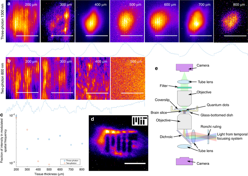
Researchers explored exactly that question. All of a sudden, wide-field (i.e., faster) imaging was possible at the cost of, essentially, nothing. Temporal focusing of a pulsed 1300 nm center-wavelength laser was used to selectively excite a 3 μm thick plane as deep as 800 μm into brain tissue – enabling 3D sectional imaging. The axial resolution was only slightly decreased from point-scanning two-photon methods while the lateral resolution remained independent of the excitation light. The study, which focused more on getting light into tissue than getting it out – which largely becomes a problem of multiply scattered light – opens up the possibility for higher-speed, deep-tissue multi-photon imaging.
With the ability to send light deep into tissue, the researchers went one step further to test the viability of three-photon optogenetics or, simply put, triggering neurons with light. The advantage of three-photon microscopy is clear – the excitation light penetrates deeper. But this work goes beyond the simple proof-of-principle that the three-photon excitation scheme is possible, and also shows that photodamage is also a multi-photon process. Using more advanced schemes and higher-power lasers, the in vivo simultaneous stimulation of hundreds of neurons deep inside of brain tissue may be possible.
Reference & Image Credit
Rowlands, C.J. et al. Wide-field three-photon excitation in biological samples. Light: Science & Applications http://dx.doi.org/10.1038/lsa.2016.255 (2017)

Leave a comment