Imaging and Diffraction

An introduction to imaging

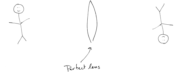
We’ve talked about light and its properties, and even microscopy, but we skipped over the basics of imaging: the way we see the world around us. An image is a visual representation of an object and, in fact, we always see images of objects, not the object themselves. When light travels to an object (let’s consider light traveling from a lightbulb to a stick figure), some of the light is absorbed (that is, it physically heats the object) and some is scattered away from the object. The scattered light goes in all directions and to see the object we must collect the scattered light and form an image. The lens in our eye does exactly that: it captures the light and directs it back together to form the image on our retina. Images can be formed of a whole scene at one moment in time (as just described) or point-by-point as is more common in microscopy.

Continue reading “Imaging and Diffraction”

Seeing with Spatial Coherence

You can’t see objects through a wall, but imagine you can see around a corner, or maybe two. While this has been demonstrated with pulsed lasers and ranging experiments, imagine instead using walls as mirrors and just looking. It may sound far-fetched, but a team led by Aristide Dogariu at CREOL at the University of Central Florida has shown that it can be done by imaging the spatial coherence of reflected light from a wall.

Continue reading “Seeing with Spatial Coherence”

Removing Noise in OCT

Seeing small objects more clearly, with less speckle.

Optical coherence tomography (OCT) is a non-invasive three-dimensional clinical imaging method that uses a coherent light source, a laser, to image different depths into tissue. However, speckle noise – an imaging artifact due to scattering and interference of coherent light – has been unavoidable and has limited the capabilities of OCT. Now, at team led by Adam de la Zerda at Stanford University has implemented a clever trick to remove speckle noise, opening up the full diagnostic potential of OCT.

Continue reading “Removing Noise in OCT”

1, 2, 3 Photons

Sometimes, more can be better.

To image deep into biological tissue, two-photon microscopy has become a standard. Two-photon microscopy uses the non-linear absorption of a fluorescent molecule to simultaneously absorb two lower-energy photons as though they were one. But what is the potential for imaging with three-photons? Continue reading “1, 2, 3 Photons”

Create a website or blog at WordPress.com

Up ↑